Week 3: 21 – 25 April 2014
Attachment in the lab of Marine Microbial Ecology under Prof. Dr Farooq Azam
Scripps Institution of Oceanography, San Diego, United States Of America
The ocean encompasses more than 70% of the planet’s surface. We have more ‘water’ around us than the ‘earth’ itself. In that ‘water’, 90% of its biomass is microbes. Did you know that in a litre of seawater, there are 10 billion viruses, 1 billion bacteria and 5 million protozoa? This reflects the abundance of microoganisms in the ocean in which the diversity can only be seen through a microscope.
Here in Professor Azam’s lab, the most routinely used microscope is the fluorescent microscopy to detect and enumerate the microbial abundance in different environmental samples. In this lab, the microscopes are used to count viruses, bacteria, microalgae and flagellates. It is also utilised to study the presence of certain organelles, growth dynamics and interaction between the species in nature and lab experimental setups.
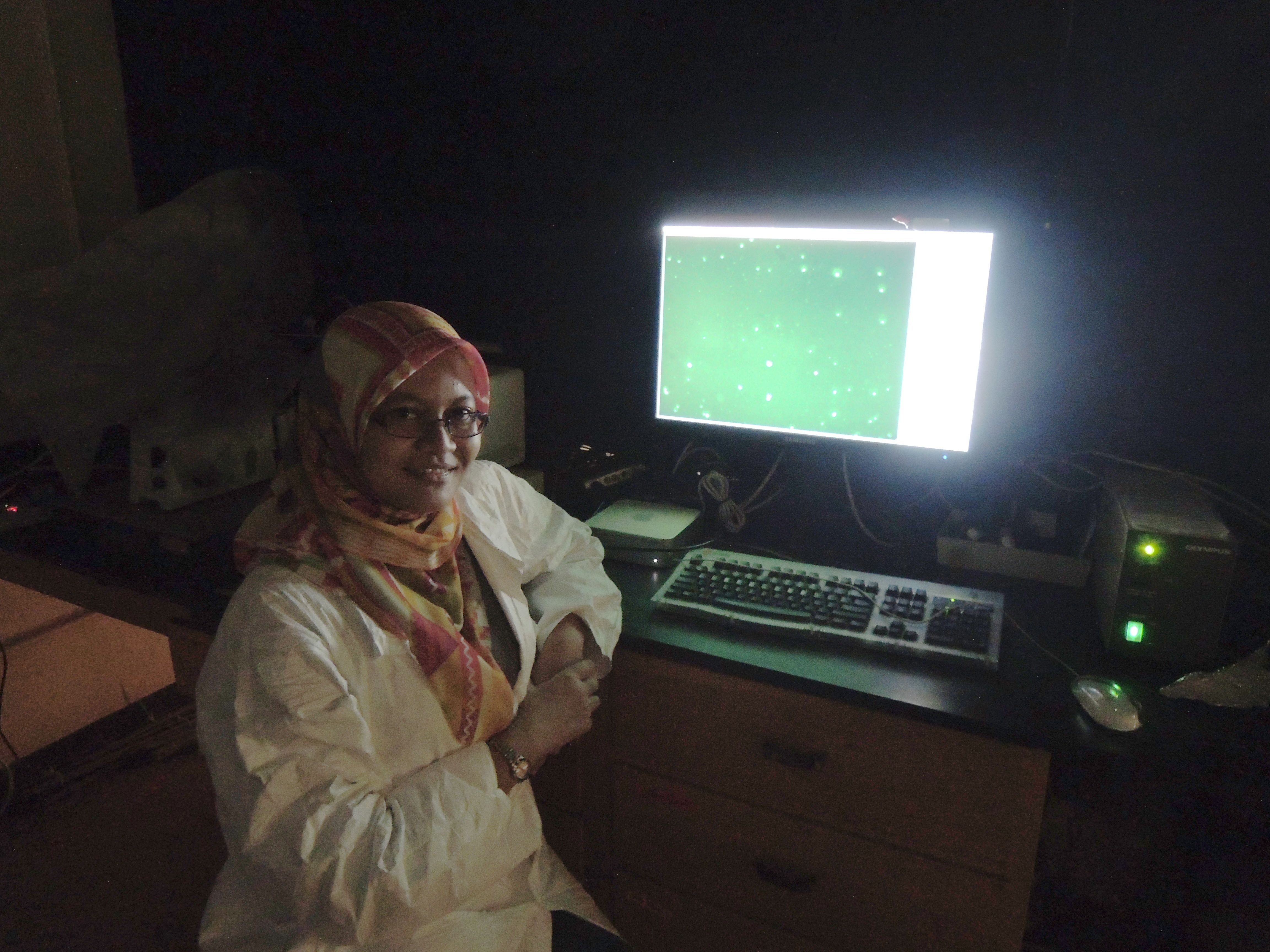
This is done through filtration of samples that are stained with different filters and fluorescent stains, respectively. Among the common dyes used are the DAPI (for nucleic acid staining & suitable for bacterial counting), SYBR Green I (nucleic acid staining & suitable for bacterial and virus counting) FM dyes (organelles staining) and NanoOrange (protein staining).
The use of different staining also enables for the detection and localisation of specific species through the use of Fluorescent
In-situ Hybridization (FISH). Using this method, certain strains of interest can be stained with fluorescently tagged molecular probe where this technique allows the sample to be differentiated from the other community. In other words, the strain of interest would exhibit different colours when compared to other species.

The lab also uses dark-field microscopy to observe and quantify the motility of the microoganisms. Using this type of microscope, the image is formed by light reflected by the specimen against dark backgrounds. Since live samples can be used, the method enables the researcher to study the interaction of different microorganisms in natural or in manipulated environment
in-situ.
Next week, the discussion will focus on confocal & atomic force microscopy. See you!